


Cranio NY Blog
CranioNY is the blog of New York Craniofacial and Plastic Surgeon, Dr. Nicholas Bastidas.
Facial Fractures
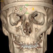

Facial Fractures
Nasal bones are the most common facial fracture, usually resulting from falls or altercations. In the first 2 weeks after the injury the bones can often be reset without making any incisions under anesthesia. Late presenting injuries require osteotomies (re-breaking the bone) to restore the normal bony architecture of your nose.
ZMC (zygomasticomaxillary complex) fractures are probably the second most common fracture. This fracture usually involves the orbital rim (lower eye socket), cheek bone (zygoma) and may also be associated with an orbital floor fracture. Displaced fractures can cause cheek asymmetry as well as sunken in appearance to the eye due to the abnormal eye socket anatomy. Unrepaired or inappropriately fixated fracture may require osteotomies (re-breaking the bone) to move the ZMC unit into anatomic position. Numbness of the cheek is quite common with these injuries.
Orbital floor fractures can cause sinking in of the eyeball and even affect the ability to move the eye normally causing double vision (diplopia). The orbital floor can be reconstructed using an implant (titanium, medpor) or a bone graft. The incision can be placed inside the eyelid to avoid visible facial scars.
Mandible fractures are also very common and may require surgery to fixate the mandible in the anatomic position using plates and screws. Frequently arch bars (wires around the teeth) are necessary to help obtain superior outcomes as well as better occlusion (ability of your teeth to meet together when you chew). Infection and poorly healed fractures may require osteotomies as well as bone grafts to properly heal the fracture.
Pierre-Robin Sequence
Pierre-Robin Sequence
A small lower jaw (mandible) can lead to abnormal tongue positioning causing a cleft palate which may obstruct the airway in some newborns. The small jaw prevents the tongue from lying forward and can be fatal if unrecognized. A craniofacial surgeon can help the problem by bringing the lower jaw forward by a process known as Mandibular distraction.
Lengthening the jaw may alleviate the obstructive sleep apnea, improving your child’s ability to breathe and feed. Tracheotomy tubes may often be prevented or removed after treatment.
Mandibular Hypoplasia
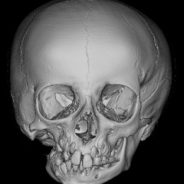

Pre-surgerical intervention
Mandibular Hypoplasia
A small lower jaw is known as mandibular hypoplasia and it can be isolated or part of a syndrome (along with other deformities). One or both sides of the jaw can be involved and treatment is aimed at lengthening the part of the jaw that is affected. This can be accomplished surgically using either bone grafts or mandibular distraction. Treatment is aimed at improving the facial aesthetics as well as the functional bite (occlusion).
Hand Trauma
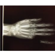

Hand Trauma
Sharp laceration, crushing injuries and avulsions can disrupt the nerves, tendons, ligaments and bones of the hand. As a fully trained plastic and reconstructive surgeon, Dr. Bastidas has significant experiences managing complex skeletal and soft tissue injuries of the hand.
Syndactyly
Syndactyly
Webbing of the fingers and toes is known as syndactyly. The involved fingers or toes can be fully fused at the level or the bone, or simply by the skin. The release of the involved structures often begins around age 12 months and typically requires a skin graft to resurface the digits.
Mobius Syndrome
Mobius Syndrome
Mobius syndrome is a congenital (birth) deformity where patients are born with facial paralysis on either one or both sides. Facial reanimation of the ability to smile can be performed using muscle transfers (temporalis or gracilis) to help bring emotion to a sometimes emotionless face.
Bells Palsy
Bell’s Palsy
Bell’s palsy is form of facial paralysis related to dysfunction of the facial nerve. Most cases are one-sided (unilateral) and the source is often unknown (idiopathic). It may be associated with lyme disease as well as with the herpes simplex virus. Treatment is aimed at restoration of facial symmetry and regaining the ability to smile. Instances involving paralysis of the eyelid may require placement of a gold weight to help protect the cornea.
Romberg’s Disease
Romberg’s Disease
Sometimes called Parry-Romberg disease, involves an autoimmune response where the subcutaneous fat of the face wastes away leaving significant contour defects. It is most common in young females, aged 5-15. Treatment strategies are developed in conjunction with a rheumatologist and may involve fat grafting and/or tissue transfer techniques to reconstruct the normal facial contour.
Hemifacial Microsomia
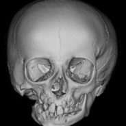

Hemifacial Microsomia
All faces are inherently asymmetric but severe asymmetry may be the result of hemifacial microsomia (HFM). HFM can be associated with orbit (eye socket), ear (microtia) and jaw malformations (mandible) as well as facial nerve palsy (facial droop). Weakness in the smile on the affected side may also be seen. Branchial clefts or remnants as well as kidney abnormalities may also be commonly associated. Surgical treatment is aimed at each involved structure, typically beginning with removal of remnants after the age of 6 months. Jaw and soft tissue abnormalities may be deferred until your child has reached school-age, around the age of five, to allow for facial growth.
OMENS classification
Microtia (small or absent ear)
Mandible Malformations
- Type 1 – vertical shortening of mandible, normal TMJ. Treatment orthodontics, mandible surgery (BSSO or distraction)
- Type 2
- Type 3 – complete absence of TMJ
Treatment around age 5: Rib Graft followed by mandibular distraction or free fibula bone graft
Soft tissue asymmteries are often treated by fat grafting
Asymmetries due to nerve palsy can be treated by Botox to the nerves on the normal side
Microtia (small or absent ear)
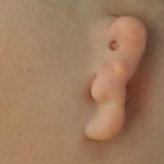

Lobule type Microtia
Microtia –(small or absent ear)
Malformation of the external ear may require complex reconstruction requiring the use of cartilage from the rib. This reconstruction is usually postponed until a minimum age of eight to allow for growth of the rib cage. A new ear framework is sculpted from the cartilages and buried underneath the skin where the new ear is to be created. Approximately three months later the “new ear” is elevated from underneath the skin and grafted to create the “normal” projection. Prosthethics are also available, however, are prone to fall off and degrade requiring expensive replacements. The cartilage sculpted ear is the preferred “permanent” reconstruction recommended by most plastic surgeons and are more resistant to infections than synthetic ears.


